Home
Our Doctor
Services
Patient Reactions
Patient Resources
Location & Hours Call 727-345-2064 DM SMILE Request a Consultation
Our Technology at Advanced Center for Cosmetic Dentistry
Our Technology

The Equipment Behind a Prosthodontist-Led Result

Published by Dr. Roberto Macedo, DDS, MS, PhD, Prosthodontist

Technology does not place a veneer or seat an implant. What it does is take the guesswork out before either one starts. Here is the equipment Advanced Center for Cosmetic Dentistry uses, what each piece changes for you, and why a prosthodontist plans a case around it.

24+ Years in practice
4.9★ Hundreds of Google reviews
In-House Master ceramists' lab
PhD Prosthodontist (DDS, MS)
Why It Matters

Plan Backwards From the Finished Smile

A prosthodontist is the dentist with specialty training in rebuilding and replacing teeth. That training changes the order of the work: the finished restoration is designed first, and everything else, the implant angle, the bone, the bite, the shade, is worked backwards from it.

That order only holds if you can see the case before you start it. Cone beam imaging shows the bone and the nerves in three dimensions. A digital scan gives a model accurate enough to design on. Our own lab then builds to that design under the same roof.

None of it is technology for its own sake. Every piece exists to answer a question earlier, while the answer is still cheap to act on.

Request a Consultation

What the Equipment Changes for You

  • Fewer surprises: the case is measured in 3D before treatment starts
  • Fewer visits: design and fabrication happen in this building
  • No impression putty: a digital scan replaces the trays
  • Lower radiation: digital sensors, not film
  • You see it too: images go on the screen beside your chair
  • One point of accountability: your restoration is not posted out
In-House Dental Lab

Our Dental Lab Is Not in Another State. It Is Down the Hall.

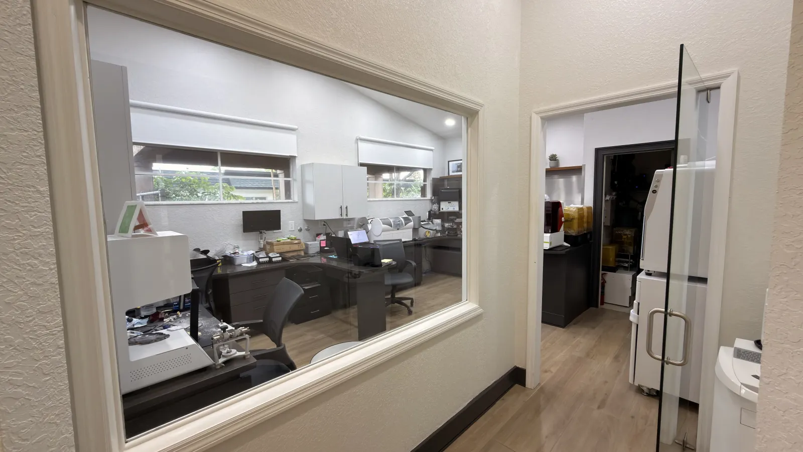
A full dental laboratory inside the practice with master ceramists on staff. Crowns, bridges, veneers, dentures, and appliances are made here rather than shipped to an outside lab, which keeps quality control in one pair of hands and cuts the wait between appointments.

Most practices design a crown and send the file to an outside laboratory they have never set foot in. Here the shade is checked against your face in daylight rather than matched from a photograph, a contour is adjusted in minutes rather than couriered back and forth, and one person owns the quality from design to polish.

01

Complete quality control

Nothing leaves the building for a stranger to interpret. Dr. Macedo and the ceramists work from the same case, in the same room.

02

Shorter waits

No shipping window in either direction. Restorations and appliances are produced in a fraction of the usual turnaround.

03

Less time without teeth

The gap between preparation and final placement shrinks, so you spend less of it in temporaries or with a space showing.

04

Everything under one roof

We rarely refer out. Planning, surgery, restoration, and fabrication happen in one location, with one person accountable.

The in-house dental laboratory seen through a glass window from the practice hallway, with workbenches, a milling unit and lab equipment
The lab, seen from the hallway
Hand finishing, 15 sec
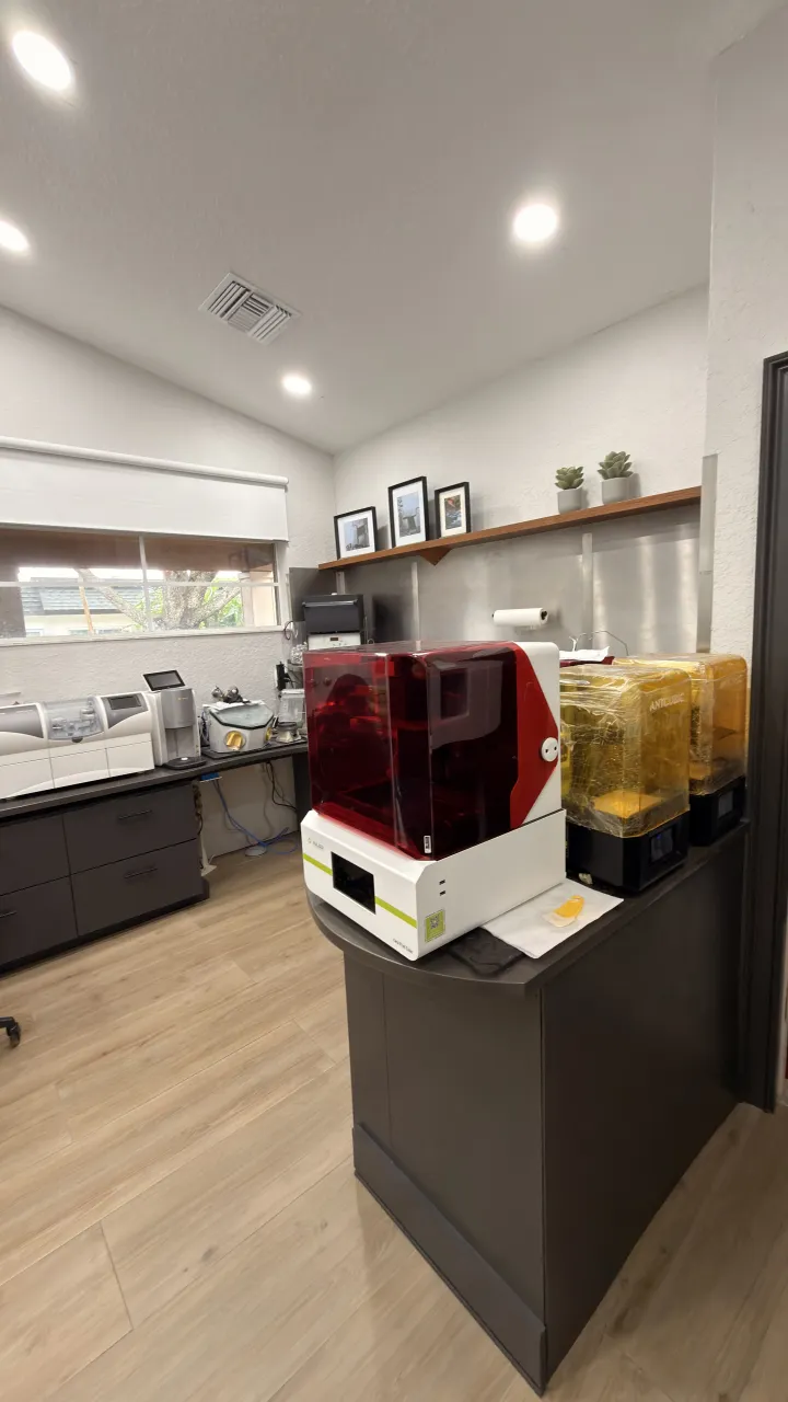
The 3D printer station inside the dental laboratory
Our 3D printer station
Imaging 01

Cone Beam 3D Imaging (CT Scanner)

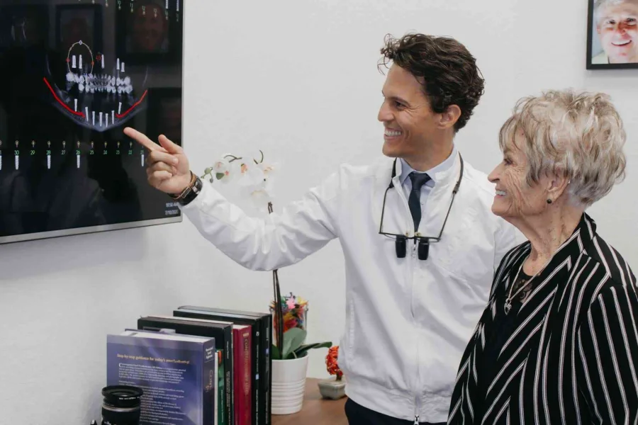
A three-dimensional reconstruction of the teeth, jaws, airway, sinuses, and surrounding facial structures. It shows bone volume, nerve position, and sinus anatomy that a flat X-ray cannot, which is what makes guided implant placement and full-arch planning possible before any surgery begins.

That measurement is what makes guided implant placement possible. Bone height and width, nerve position, and sinus floor are known before the first incision, not discovered during it.

Cone beam gives a more complete view of the mouth and its supporting structures than digital X-rays alone. Both are used, for different questions.

Dr. Roberto Macedo reviewing a 3D implant plan on a wall monitor with a patient

What we use a cone beam scan for

  • Plan dental implant placement
  • Evaluate the jaws and face
  • View the head and neck as a whole
  • Diagnose decay and other dental problems
  • Detect endodontic problems and plan root canal therapy
  • Analyze dental and facial trauma
  • Plan and track orthodontic treatment
  • Visualize abnormal or unerupted teeth
  • Assess a TMJ disorder
Imaging 02

Digital X-Rays

Sensor-based radiography that appears on a chairside monitor within seconds, with no film and no developing chemicals. Digital X-rays produce 80 to 90 percent less radiation than film radiography and are used to find decay between the teeth, bone loss, abscesses, fractured fillings, and infection in the nerve.

80 to 90% less radiation than film radiography

What a digital X-ray finds

  • Decay between the teeth or in the roots
  • Bone loss around the teeth
  • Cysts and abscesses
  • Fractures in existing fillings
  • Infection in the tooth nerve
  • Developmental abnormalities
  • Improper root positioning
  • Tumors

Digital X-rays are also standard in planning oral surgery, including implant placement and root canal therapy.

Chairside

Three Tools You Will Actually Notice

The imaging happens around you. These three happen to you, and each one changes the appointment itself.

Intraoral Scanner

Impressions without the putty

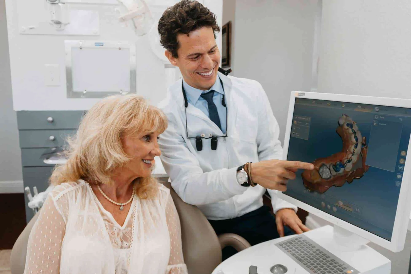
A digital impression system that replaces trays of impression putty. It captures a precise 3D model of the teeth for crowns, veneers, and clear aligners, can be stopped and restarted at any moment, and is far easier for patients with a strong gag reflex.

Used for crowns, veneers, and Invisalign aligners.

Intraoral Camera

You see what the dentist sees

A pen-sized camera that reaches areas the eye cannot see and projects detailed color images onto a screen beside you. It sharpens the diagnosis of decay, gum disease, and cracked teeth, and it lets you see exactly what the dentist sees rather than take it on trust.

Used for diagnosis, patient education, and records.

Magnification Loupes

1.5x to 6x beyond normal vision

Binocular surgical telescopes worn like eyeglasses that magnify the working field 1.5 to 6 times beyond normal vision. The extra detail catches early problems while they are still small and allows margins and fine finishing work to be cut with far greater precision.

Worn for exams, restorative work, and cosmetic finishing.

The Workflow

Digital Dentistry and Same-Day Restorations

The digital chain that connects scanning, design, and fabrication. A tooth is scanned, the restoration is designed on screen, and it is produced in our own laboratory, so some cases are finished in a single day instead of across weeks of temporaries.

The same chain is what lets you preview a smile before anything is cut. A veneer case is designed on screen and shown to you first, so the approval happens before the drill, not after.

Whether your case is single-visit depends on the tooth, the bite, and the shade. We confirm the sequence at your consultation instead of promising it up front.

How a same-day crown works
1

Scan

The intraoral scanner captures a 3D model of the tooth and the bite, with no impression material.

2

Design

The restoration is designed on screen against your existing teeth, then reviewed with you.

3

Fabricate

Our in-house lab produces it. No courier, no outside technician interpreting the file.

4

Fit and finish

Bite, contour, and shade are adjusted in the chair, with the ceramists a room away if anything needs changing.

Common Questions

Our Technology, Answered

What dental technology does Advanced Center for Cosmetic Dentistry use?

Seven pieces of equipment carry most of the clinical work: a cone beam 3D CT scanner, digital X-rays, an intraoral scanner for digital impressions, an intraoral camera, magnification loupes, the digital design workflow behind same-day restorations, and a full dental laboratory inside the practice with master ceramists on staff.

Do you have a dental lab on site?

Yes. Crowns, bridges, veneers, dentures, and appliances are made in our own laboratory by our in-house master ceramists rather than posted to an outside lab. It means one set of hands owns the quality, the shade can be checked against your face in the same building, and the wait between appointments is much shorter.

Are digital X-rays safer than film X-rays?

Digital X-rays produce 80 to 90 percent less radiation than film radiography. They also use no developing chemicals and appear on a monitor within seconds, so the image can be reviewed with you in the same appointment rather than at the next one.

What is a cone beam CT scan and why would I need one?

A cone beam scan builds a three-dimensional model of your teeth, jaws, and surrounding structures instead of the flat picture a standard X-ray gives. Dr. Macedo uses it to measure bone volume before an implant, to locate nerves and sinuses, to plan root canal therapy, to assess trauma or a TMJ problem, and to see the head and neck as one connected structure.

Do I still have to bite into impression putty?

For most cases, no. An intraoral scanner captures a digital impression instead. There is nothing to set or swallow, the scan can be stopped and restarted at any point, and the resulting model is precise enough to design a crown or a set of aligners from. Patients who gag easily usually find it the single biggest improvement in the whole visit.

Can you make a crown in one day?

Some cases can be designed and produced on the same day, because the scanning, design, and fabrication all happen in this building. Whether yours is a single-visit case depends on the tooth, the bite, and the shade match. Dr. Macedo confirms the sequence at your consultation rather than promising it in advance.

What is an intraoral camera used for?

It photographs areas of your mouth that are difficult to examine by eye and projects them onto a screen you can see. It supports a more accurate diagnosis of decay, gum disease, and tooth damage, it records the result after treatment, and the images can be sent to another specialist or to your insurance provider when needed.

Why does the dentist wear magnifying loupes?

Loupes are binocular surgical telescopes that magnify the field 1.5 to 6 times. The detail they reveal means smaller problems are found earlier, when treatment is less invasive and less expensive, and it allows preparation margins and cosmetic finishing to be cut with a precision that is not achievable by naked eye.

St. Petersburg, FL

See It Before You Commit to It

A consultation starts with the imaging, not the sales pitch. You see your own scans, your own options, and the sequence, before you decide anything.